| dc.contributor.author | Tunalı, Varol | |
| dc.contributor.author | Harman, Mehmet | |
| dc.contributor.author | Cavus, Ibrahim | |
| dc.contributor.author | Gunduz, Cumhur | |
| dc.contributor.author | Ozbilgin, Ahmet | |
| dc.contributor.author | Turgay, Nevin | |
| dc.date.accessioned | 2020-11-20T14:30:01Z | |
| dc.date.available | 2020-11-20T14:30:01Z | |
| dc.date.issued | 2020 | |
| dc.identifier.issn | 1230-2821 | |
| dc.identifier.issn | 1896-1851 | |
| dc.identifier.uri | https://doi.org/10.1007/s11686-020-00285-0 | |
| dc.identifier.uri | https://hdl.handle.net/20.500.12809/318 | |
| dc.description | WOS: 000573738000003 | en_US |
| dc.description | PubMed ID: 32996014 | en_US |
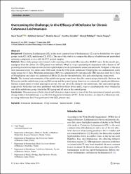
| dc.description.abstract | Background Cutaneous Leishmaniasis (CL) is the most common form of leishmaniasis. CL can be divided into two major groups: acute CL (ACL) and chronic CL (CCL). The aim of this study is to compare the efficacy of miltefosin and pentavalent antimony compoundsin vivowith the CCL patient samples. Materials Three study groups were formed, each consisting of five male Mus musculus (Balb/C) mice. In this model, promastigotes from the culture of a CCL patient were utilized. 100 mu LL. tropicapromastigote suspension with a density of 10(8)promastigotes/ml were injected into the hint-right footpad of each experimental animal intradermally. Footpads of the mice were measured every two weeks until 24th week. From the 13th week, miltefosin 50 mg/kg/day was administered orally using gavage for 21 days, Meglumin antimoniate (MA) was administered by intramuscular (IM) injection daily for 21 days at 50 mg/kg/day and saline was administered IM for 21 days for the miltefosine, MA and control group, respectively. Results The footpad measurements of the miltefosine group were lower than the control group statistically. Between the MA group and the miltefosine group and MA group and the control group, there was no statistically significant difference. Giemsa stained slides revealed amastigotes in one, two and all of the slides for the miltefosine, MA and control group, respectively. Molecular tests were performed with the Rotor-Gene device andL. tropicaconsistent peaks were obtained in one of the miltefosine group, four in the MA group and all mice in the control group. Conclusions Demonstration of both clinical and laboratory improvement in four of the five experimental animals provides strong evidence that miltefosine is an effective drug in the treatment of CCL. In the literature, no clinical or laboratory studies using miltefosine have been performed with CCL patients only. | en_US |
| dc.description.sponsorship | Research Funds of Ege University [17-TIP-017] | en_US |
| dc.description.sponsorship | This project was supported by Research Funds of Ege University with the Project no: 17-TIP-017. | en_US |
| dc.item-language.iso | eng | en_US |
| dc.publisher | Springer International Publishing Ag | en_US |
| dc.item-rights | info:eu-repo/semantics/openAccess | en_US |
| dc.subject | Cutaneous Leishmaniasis | en_US |
| dc.subject | Drug Resistance | en_US |
| dc.subject | Animal Model | en_US |
| dc.subject | Turkey | en_US |
| dc.title | Overcoming the Challenge; In VivoEfficacy of Miltefosine for Chronic Cutaneous Leishmaniasis | en_US |
| dc.item-type | article | en_US |
| dc.contributor.department | MÜ, Tıp Fakültesi, Cerrahi Tıp Bilimleri | en_US |
| dc.contributor.institutionauthor | Tunalı, Varol | |
| dc.identifier.doi | 10.1007/s11686-020-00285-0 | |
| dc.relation.journal | Acta Parasitologica | en_US |
| dc.relation.publicationcategory | Makale - Uluslararası Hakemli Dergi - Kurum Öğretim Elemanı | en_US |


