| dc.contributor.author | Çetin, Sedat | |
| dc.contributor.author | Yur, Fatmagül | |
| dc.contributor.author | Taşpınar, Mehmet | |
| dc.contributor.author | Yüksek, Veysel | |
| dc.date.accessioned | 2020-11-20T14:41:40Z | |
| dc.date.available | 2020-11-20T14:41:40Z | |
| dc.date.issued | 2019 | |
| dc.identifier.issn | 0015-4725 | |
| dc.identifier.issn | 2253-4083 | |
| dc.identifier.uri | https://hdl.handle.net/20.500.12809/963 | |
| dc.description | WOS: 000482767500008 | en_US |
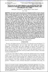
| dc.description.abstract | The present study was planned to investigate the effects of some minerals (MgCl2, Na2SeO3, AlCl3 CaCl2) on the expression and translocation of certain apoptotic markers in NaF-administered (at the rate of IC50) rat renal epithelial (NRK-52E) and human osteoblast (hFOB 1.19) cell lines. The NaF IC50 and the non-toxic mineral doses were determined by 3-(4,5-dimethylthiazol-2-yl)-2,5-diphenyltetrazolium bromide (MTT) assay. For the biochemical analysis, the cells were collected by trypsin treatment following a 24-hour incubation period, the cells were broken up by the freeze/thaw method, and the analysis was conducted. Caspase 9, 8, and 3 levels and gene expression, and M30 and 8-OHdG levels were determined. Target gene DNAs were propagated with the real time-PCR method. In the MTT studies, it was determined that the cell proliferation in rat renal epithelial cells (NRK-52E) treated with NaF+minerals was higher than that of all the NaF-treated groups and in the human osteoblast cells (hFOB 1.19), the cell proliferation was higher than in all the NaF-treated groups except for the MgCl2 group. The reason why the NaF administration in the NRK-52E cells resulted in an average of a 2-fold decrease in caspase 3 expression compared to the control group could be attributed to the apoptotic effect of NaF based on the time we obtained the RNA. However, based on this time, when the results are assessed based on the NaF and other mineral groups, the NaF-induced cytotoxic apoptosis might have used a pathway other than the apoptotic pathway. Thus, it is considered that minerals could usually prevent NaF-induced apoptosis by a synergistic mechanism due to the ionic character of NaF. NaF+mineral administration protected the NRK-52E cells from apoptosis. In the osteoblasts, on the other hand, it was concluded that NaF+mineral administration may be useful since it inhibits increased apoptosis. | en_US |
| dc.description.sponsorship | Van Yuzuncu Yil University Research Projects Directorate [2015-SBE-D200] | en_US |
| dc.description.sponsorship | This research was approved by the Van Yuzuncu Yil University Research Projects Directorate (Project no: 2015-SBE-D200) and based on a dissertation by Fatmagul Yur, | en_US |
| dc.item-language.iso | eng | en_US |
| dc.publisher | Int Soc Fluoride Research | en_US |
| dc.item-rights | info:eu-repo/semantics/openAccess | en_US |
| dc.subject | Apoptosis | en_US |
| dc.subject | Cell Culture | en_US |
| dc.subject | Minerals | en_US |
| dc.subject | Naf | en_US |
| dc.subject | Real Time-PCR | en_US |
| dc.title | THE EFFECTS OF SOME MINERALS ON APOPTOSIS AND DNA DAMAGE IN SODIUM FLUORIDE-ADMINISTERED RENAL AND OSTEOBLAST CELL LINES | en_US |
| dc.item-type | article | en_US |
| dc.contributor.department | MÜ, Fethiye Sağlık Bilimleri Fakültesi, Beslenme Ve Diyetetik Bölümü | en_US |
| dc.contributor.institutionauthor | Yur, Fatmagül | |
| dc.identifier.volume | 52 | en_US |
| dc.identifier.issue | 3 | en_US |
| dc.identifier.startpage | 362 | en_US |
| dc.identifier.endpage | 378 | en_US |
| dc.relation.journal | Fluoride | en_US |
| dc.relation.publicationcategory | Makale - Uluslararası Hakemli Dergi - Kurum Öğretim Elemanı | en_US |


